Histopathological patterns identified in skin biopsies with tegumentary leishmaniasis in Colombia.
DOI:
https://doi.org/10.29176/2590843X.2070Keywords:
Granuloma, Hematoxylin-eosin, Inflammation, Leishmania, Lymphoplasmacytic infiltrate, Mucocutaneous leishmaniasis, Parasite load, UlcerAbstract
Introduction: Tegumentary leishmaniasis (TL) is a neglected disease that affects the skin and mucous membranes. Its detection poses a challenge due to the lack of a gold standard for diagnosis.
Objective: To describe the histopathological patterns associated with a group of samples from patients with tegumentary leishmaniasis.
Methods: This descriptive study was conducted through histopathological evaluation using hematoxylin-eosin staining on 43 paraffin-embedded samples of chronic tegumentary leishmaniasis, categorized into three groups: moderate to high parasite load (n=15), low load (n=15), and chronic granulomatous inflammation (n=13).
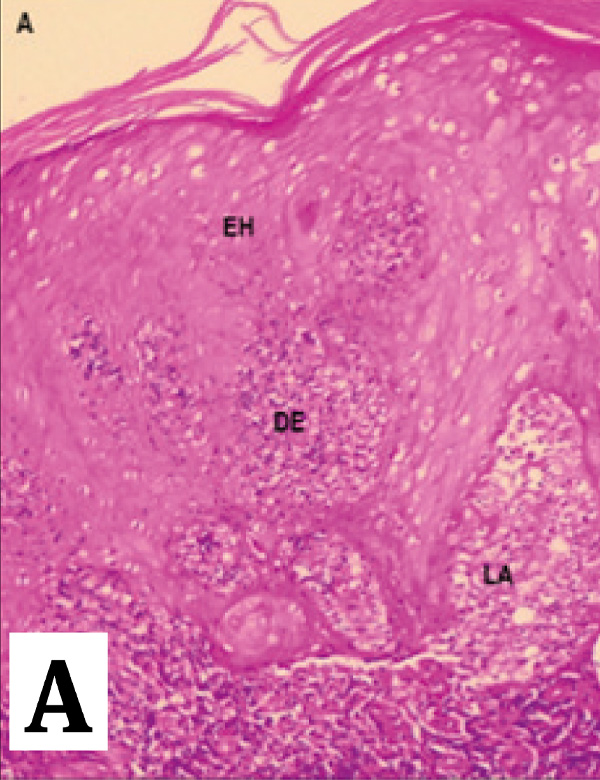
Results: Epidermal hyperplasia was observed in 34.9% of cases, and ulcers were present in 62.8% of cases. A predominant lymphoplasmacytic infiltrate was found in 67.4% of samples, while a histiocytic infiltrate was observed in 90.7% of samples. In samples with no detectable parasite load, the histiocytic infiltrate was present in 94.8% of cases. In samples with low parasite load, the histiocytic infiltrate was significant in 55.5% of cases, and 86.6% of the samples with moderate to high parasite load exhibited this histopathological pattern.
Conclusions: Lymphoplasmacytic and histiocytic infiltrates are consistent histopathological patterns, even in samples with low or absent parasite load.
Author Biographies
Lucero Katherine Aristizábal-Parra , Universidad CES, Medellín, Colombia
Instituto Colombiano de Medicina Tropical, Universidad CES, Medellín, Colombia.
Juan Pablo Ospina-Gómez, Universidad de Antioquia, Medellín, Colombia
Centro de Investigaciones en Dermatología - CIDERM, Facultad de Medicina, Universidad de Antioquia, Medellín, Colombia.
Héctor Serrano-Coll, Universidad CES, Medellín, Colombia
Instituto Colombiano de Medicina Tropical, Universidad CES, Medellín, Colombia.
References
Gabriel Á, Valério-Bolas A, Palma-Marques J, Mourata-Gonçalves P, Ruas P, Dias-Guerreiro T, et al. Cutaneous Leishmaniasis: The Complexity of Host’s Effective Immune Response against a Polymorphic Parasitic Disease. J Immunol Res. 1 de diciembre de 2019;2603730.
Daga MK, Rohatgi I, Mishra R. Leishmaniasis. Indian J Crit Care Med Peer-Rev Off Publ Indian Soc Crit Care Med. mayo de 2021;25(Suppl 2):S166-70.
E. Aronson N, J. Magill A. Leishmaniasis. En: Hunter’s Tropical Medicine and Emerging Infectious Diseases. 10th ed. ELSEVIER; 2020. p. 776-98.
Torres-Guerrero E, Quintanilla-Cedillo MR, Ruiz-Esmenjaud J, Arenas R. Leishmaniasis: a review. F1000Research. 26 de mayo de 2017;6:750.
Zambrano PI. Protocolo de vigilancia en salud pública Leishmaniasis. 6 de marzo de 2020;(04):16.
Chivatá NJA. Informe de evento Leishmaniasis cutánea, mucosa y visceral, Colombia, 2018. 31 de mayo de 2019;(04):28.
Organización Panamericana de la Salud O mundial de la salud. Leishmaniasis, informe epidemiológico de las Américas [Internet]. 2022. Disponible en: https://iris.paho.org/bitstream/handle/10665.2/56833/OPASCDEVT220021_spa.pdf?sequence=1&isAllowed=y
M P, Jr R, Ga R, Ce PF, K OA, An SME, et al. Interventions for American cutaneous and mucocutaneous leishmaniasis. Cochrane Database Syst Rev [Internet]. 27 de agosto de 2020 [citado 13 de marzo de 2022];8(8). Disponible en: https://pubmed.cesproxy.elogim.com/32853410/
González K, Diaz R, Ferreira AF, García V, Paz H, Calzada JE, et al. Histopathological characteristics of cutaneous lesions caused by Leishmania Viannia panamensis in Panama. Rev Inst Med Trop Sao Paulo. 2018;60:e8.
Boussoffara T, Boubaker MS, Ben Ahmed M, Mokni M, Guizani I, Ben Salah A, et al. Histological and immunological differences between zoonotic cutaneous leishmaniasis due to Leishmania major and sporadic cutaneous leishmaniasis due to Leishmania infantum. Parasite Paris Fr. 2019;26:9.
Wijesinghe H, Gunathilaka N, Semege S, Pathirana N, Manamperi N, de Silva C, et al. Histopathology of Cutaneous Leishmaniasis Caused by Leishmania donovani in Sri Lanka. BioMed Res Int. 2020;2020:4926819.
Serrano-Coll H, Cardona-Castro N, Ramos AP, Llanos-Cuentas A. Innate immune response: ally or enemy in cutaneous leishmaniasis? Pathog Dis. 8 de junio de 2021;79(5):ftab028.
Abbas A, Lichtman A, Pillai S. Cellular and molecular immunology [Internet]. Elsevier/Saunders; 2015 [citado 14 de enero de 2017]. Disponible en: https://www.ncbi.nlm.nih.gov/nlmcatalog/101630458
Carvalho AM, Bacellar O, Carvalho EM. Protection and Pathology in Leishmania braziliensis Infection. Pathog Basel Switz. 14 de abril de 2022;11(4):466.
How to Cite
Downloads
Downloads
Published
How to Cite
Issue
Section
License
Copyright (c) 2025 Lucero Katherine Aristizábal-Parra , Juan Pablo Ospina-Gómez, Héctor Serrano-Coll

This work is licensed under a Creative Commons Attribution-NonCommercial-ShareAlike 4.0 International License.
| Article metrics | |
|---|---|
| Abstract views | |
| Galley vies | |
| PDF Views | |
| HTML views | |
| Other views | |