Langerhans' cell histiocytosis: An update based on a real case. Part 2
DOI:
https://doi.org/10.29176/30284163.1961Keywords:
Congenital self-healing reticulohistiocytosis (Hashimoto-Pritzker disease), Cutaneous neo plasms, Langerhans cell histiocytosis, Non-Langerhans cell histiocytosisAbstract
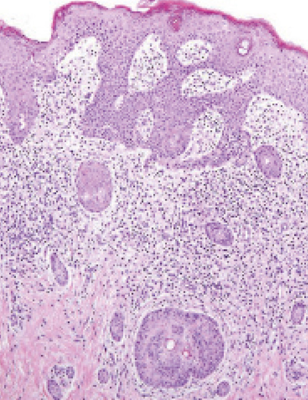
Histiocytoses are rare disorders characterized by the accumulation of dendritic cells or macrophages in various
organs, with a predilection for the skin and bone (1). Its clinical behavior varies from mild to life-threatening
forms(2). The first classification of histiocytosis in 1987 (3) consisted of three categories: Langerhans cell (LCH),
non-LC-related (HCNL), and malignant histiocytosis (MH). These inflammatory neoplasms can present in limited,
diffuse forms or with multiorgan involvement (4), and manifest in multiple ways and mimic other diseases, which
is why they represent a diagnostic challenge for the clinic.
Author Biographies
Mario Zavala-Mena, Universidad de Santiago de Chile.
Residente de Dermatología, Universidad de Santiago de Chile.
Isabel Jimeno-Ortega, Universidad de Santiago de Chile.
Residente de Dermatología, Universidad de Santiago de Chile.
Gabriel Aedo-Inostroza, Universidad de Santiago de Chile.
Dermatólogo, Universidad de Santiago de Chile.
How to Cite
Downloads
Downloads
Published
How to Cite
Issue
Section
License
Copyright (c) 2024 Mario Zavala-Mena, Isabel Jimeno-Ortega, Gabriel Aedo-Inostroza

This work is licensed under a Creative Commons Attribution-NonCommercial-ShareAlike 4.0 International License.