Skin lesions in pediatrics: highlighting an entity with distinctive histopathology
DOI:
https://doi.org/10.29176/30284163.2008Keywords:
Histiocytosis, Skin diseases, Xanthogranuloma, juvenileAbstract
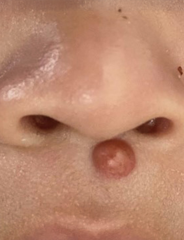
Juvenile xanthogranuloma (JXG) is the most common form of non-Langerhans cell histiocytosis, a rare group of disorders typically seen in pediatric patients, especially in children under five years old. JXG most commonly presents as localized cutaneous lesions, characterized by yellowish papules or nodules, often found on the head and neck. In the majority of cases, the condition is benign and resolves spontaneously within three to seven years, occasionally leaving behind residual hyperpigmentation or atrophy. However, in some instances, JXG can involve multiple organ systems, potentially impacting the patient’s health or life. In such cases, urgent and personalized treatment is required.
Author Biographies
Santiago Beuth Ruiz, Universidad de Antioquia. Medellín, Colombia
Médico residente de Dermatología, Universidad de Antioquia.
María Alejandra Correa-Trujillo, Universidad de Antioquia, Medellín, Colombia
Estudiante de Medicina, Universidad de Antioquia.
Juan David Ruiz-Restrepo, Universidad de Antioquia, Medellín, Colombia
Dermatopatólogo y docente de dermatopatología, Universidad de Antioquia; dermatopatólogo, Fundación Colombiana de Cancerología-Clínica Vida
Juan Pablo Ospina-Gómez, Universidad de Antioquia, Medellín, Colombia
Dermatopatólogo y docente de dermatopatología, Universidad de Antioquia; dermatopatólogo, Fundación Colombiana de Cancerología-Clínica Vida
María Natalia Mejía-Barreneche, Universidad de Antioquia, Medellín, Colombia
Dermatóloga y docente de Dermatología, Universidad de Antioquia; Dermatóloga, hospital infantil San Vicente Fundación.
References
Hernández-San Martín MJ, Vargas-Mora P, Aranibar L. Xantogranuloma juvenil: una entidad con amplio espectro clínico, Actas DermoSifiliográficas. 2020;111(9):725-33. https://doi.org/10.1016/j.ad.2020.07.004
Collie JS, Harper CD, Fillman EP. Juvenile Xanthogranuloma [Internet]. StatPearls. Treasure Island (FL): StatPearls Publishing; 2024. Disponible en: https://pubmed.ncbi.nlm.nih.gov/30252359/
McClain KL, Bigenwald C, Collin M, Haroche J, Marsh RA, Merad M, et al. Histiocytic disorders. Nat Rev Dis Primers. 2021;7(1):73. https://doi.org/10.1038/s41572-021-00307-9
Warren T. Goodman, Terry L. Barrett. Histiocitosis. En: Bolognia JL, Schaffer JV, Cerroni L (editores). Dermatología. 4.a edición. Madrid: Elsevier; 2019. p.1614-33.
Miraglia E, Laghi A, Moramarco A, Giustini S. Juvenile xanthogranuloma in neurofibromatosis type 1. Prevalence and possible correlation with lymphoproliferative diseases: experience of a single center and review of the literature. Clin Ter. 2022;173(4):353-5. https://doi.org/10.7417/CT.2022.2445
Paxton, CN, O’Malley DP, Bellizzi AM, Alkapalan D, Fedoriw Y, Hornick JL, et al. Genetic evaluation of juvenile xanthogranuloma: genomic abnormalities are uncommon in solitary lesions, advanced cases may show more complexity. Mod Pathol. 2017;30(9):1234-40. https://doi.org/10.1038/modpathol.2017.50
So N, Liu R, Hogeling M. Juvenile xanthogranulomas: Examining single, multiple, and extracutaneous presentations. Pediatr Dermatol. 2020;37(4):637-44. https://doi.org/10.1111/pde.14174
Samuelov L, Kinori M, Chamlin SL, Wagner A, Kenner-Bell BM, Paller AS, et al. Risk of intraocular and other extracutaneous involvement in patients with cutaneous juvenile xanthogranuloma. Pediatr Dermatol. 2018;35(3):329-35. https://doi.org/10.1111/pde.13437
Weiss VL, Brock JW, Cajaiba MM. Juvenile Xanthogranuloma: An Unusual Cause of Intratesticular Mass in Childhood. Urology. 2014;83(5):1109-12. https://doi.org/10.1016/j.urology.2013.12.034
Tamir I, Davir R, Fellig Y, Weintraub M, Constantini S, Spektor S. Solitary juvenile xanthogranuloma mimicking intracranial tumor in children. J Clin Neurosci. 2013;20(1):183-8. https://doi.org/10.1016/j.jocn.2012.05.019
Peruilh-Bagolini L, Silva-Astorga M, Hernández San Martín MJ, Manoli MS, Papageorgiou C, Apalla Z, et al. Dermoscopy of Juvenile Xanthogranuloma. Dermatology. 2021;237(6):946-51. https://doi.org/10.1159/000510265
Fraitag S, Barete S. 91 – Histiocytoses En: Bolognia JL, Schaffer JV, Cerroni L (editores). Dermatology 5.a edición. EUA: Elsevier; 2024. p. 1629-48.
Salari B, Dehner LP. Juvenile and adult xanthogranuloma: A 30-year single-center experience and review of the disorder and its relationship to other histiocytoses. Ann Diagn Pathol. 2022;58:151940. https://doi.org/10.1016/j.anndiagpath.2022.151940
Stover DG, Alapati S, Regueira O, Turner C, Whitlock JA. Treatment of juvenile xanthogranuloma. Pediatric Blood Cancer. 2008;51(1):130-3. https://doi.org/10.1002/pbc.21523
Cournoyer E, Ferrell J, Sharp S, Ray A, Jordan M, Dandoy C, et al. Dabrafenib and trametinib in Langerhans cell histiocytosis and other histiocytic disorders. Haematologica. 2024 ;109(4):1137-48. https://doi.org/10.3324/haematol.2023.283295
Lian H, Wei A, He L, Yang Y, Ma H, Zhang L, et al. Clinical Analysis of Pediatric Systemic Juvenile Xanthogranulomas: A Retrospective Single-Center Study. Front Pediatr. 2021;9:672547. https://doi.org/10.3389/fped.2021.672547
Maeda M, Morimoto A, Shioda Y, Asano T, Koga Y, Nakazawa Y, et al. Long-term outcomes of children with extracutaneous juvenile xanthogranulomas in Japan. Pediatr Blood Cancer. 2020;67
How to Cite
Downloads
Downloads
Published
How to Cite
Issue
Section
License
Copyright (c) 2025 Santiago Beuth Ruiz, María Alejandra Correa-Trujillo, Juan David Ruiz-Restrepo, Juan Pablo Ospina-Gómez, María Natalia Mejía-Barreneche

This work is licensed under a Creative Commons Attribution-NonCommercial-ShareAlike 4.0 International License.