Inflammatory tinea: kerion celsi. Presentation of a case
DOI:
https://doi.org/10.29176/2590843X.1816Keywords:
Tinea capitis, kerion, dermatophytesAbstract
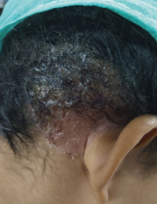
Tinea capitis is defined as an infection or parasitism of the hair, scalp, eyebrows, and eyelashes. The clinical presentation is variable, depending on the type of hair invasion, the level of resistance, and the degree of host inflammatory response. The most severe reaction pattern is known as kerion Celsi, caused mainly by zoophilic dermatophytes, Microsporum canis and Trichophyton tonsurans. It begins as a dry ringworm, which later presents with erythema, inflammation, pustules, and honey-colored crusts, from which abundant pus drains. Due to this clinical appearance, it takes the name kerion, which means “honeycomb”. It can be associated with regional lymphadenopathy, fever, malaise, local pain, and often resolves, leaving an area of scarring alopecia. Early diagnosis and timely treatment are important to avoid these sequelae.
Author Biographies
Marilyn Dayana Rivero-Bermúdez, Universidad de Carabobo. Venezuela
Residente de segundo año del posgrado de Dermatología, Universidad de Carabobo. Venezuela
Elianny Andazora, Universidad de Carabobo. Venezuela
Residente de segundo año del posgrado de Dermatología, Universidad de Carabobo. Venezuela
Sandra Vivas, Universidad de Carabobo. Venezuela
Médica internista y dermatóloga. Jefa, del Servicio de Dermatología, Ciudad Hospitalaria “Dr. Enrique Tejera”. Coordinadora académica, programa de posgrado de Dermatología, Universidad de Carabobo. Magíster en Investigación Educativa. Venezuela.
References
Bonifaz A. Micología medica básica. 4 ed. Mexico: Mcgraw-Hill; 2012.
John AM, Schwartz RA, Janniger CK. The kerion: an angry tinea capitis. Int J Dermatol. 2018;57(1):3–9. http://dx.doi.org/10.1111/ijd.13423.
Santos PE, Córdoba S, Rodero LL, Carrillo-Muñoz AJ, Lopardo HA. Tinea capitis. Experiencia de 2 años en un hospital de pediatría de Buenos Aires, Argentina. Rev Iberoam Micol. 2010;27(2):104–6. https://linkinghub.elsevier.com/retrieve/pii/S1130140610000124.
Hay RJ. Tinea Capitis: Current Status. Mycopathologia. 2017;182(1-2):87-93. https://doi.org/10.1007/s11046-016-0058-8.
Vargas N, Ayala GA, Franco C, Pablo J, Caicedo M, Rojas JP. Tiña Capitis en niños Tinea capitis en niños. Rev Chil Pediatr. 2020;91(5):773-783. http://dx.doi.org/10.32641/rchped.vi91i5.1345.
Wei S, Wang H, Li A, Yuan C. Kerion Celsi caused by Microsporum gypseum in a Chinese child, a case report. Medicine 2022;101:13 http://dx.doi.org/10.1097/MD.0000000000028936.
Arrazola J, Isa R, Torres E, Arenas R. Tinea capitis. Dermoscopic findings in 37 patients. Rev Iberoam Micol. 2015;32(4):242–6. http://dx.doi.org/10.1016/j.riam.2014.09.002
Messina F, Walker L, Romero M de LM, Arechavala AI, Negroni R, Depardo R, et al. Tinea capitis: aspectos clínicos y alternativas terapéuticas. Rev Argent Microbiol. 2021;53(4):309–13. http://dx.doi.org/10.1016/j.ram.2021.01.004.
Chan YC, Friedlander SF. Therapeutic options in the treatment of tinea capitis. Expert Opinion on Pharmacotherapy. 2004;5(2):219–27. http://dx.doi.org/10.1517/eoph.5.2.219.26472.
Rebollo N, López-Barcenas AP, Arenas R. Tiña de la cabeza. Actas Dermosifiliogr. 2008;99(2):91–100. http://www.actasdermo.org/es-tina-cabeza-articulo-S0001731008746301.
How to Cite
Downloads
Downloads
Published
How to Cite
Issue
Section
License
Copyright (c) 2024 Marilyn Dayana Rivero-Bermúdez, Elianny del Carmen Andazora-González, Sandra Carlina Vivas Toro

This work is licensed under a Creative Commons Attribution-NonCommercial-ShareAlike 4.0 International License.
| Article metrics | |
|---|---|
| Abstract views | |
| Galley vies | |
| PDF Views | |
| HTML views | |
| Other views | |