Dermoscopic characteristics of longitudinal nail groove secondary to digital myxoid cyst
DOI:
https://doi.org/10.29176/2590843X.1830Keywords:
Digital myxoid cyst, Longitudinal nail groove, Nail dystrophyAbstract
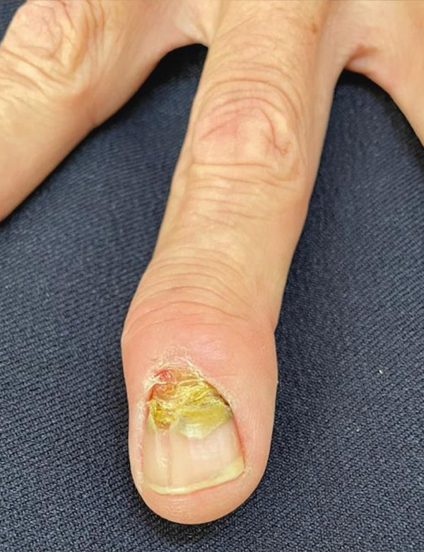
The longitudinal nail fold is a type of onychodystrophy that typically appears as a groove beginning at the proximal fold towards the distal edge. Its etiology is unknown, although it has been associated with the presence of tumors that directly damage the matrix. Myxoid cysts, despite being a frequent entity in dermatology, it is necessary to consider other diagnoses associated with nail pathology since these are not visible in early stages. Dermoscopy can be a support tool for etiology identification of the dystrophy. We present a case with clinical and dermoscopic diagnosis of longitudinal nail groove secondary to a digital myxoid cyst.
Author Biographies
Andrea Isabel Méndez-Juárez, Secretaría de Salud, Jalisco, México
Residente de segundo año de Dermatología, Instituto Dermatológico de Jalisco Dr. José Barba Rubio, Secretaría de Salud, Jalisco, México.
Enrique Adolfo Orozco-Yee, Secretaría de Salud, Jalisco, México
Dermatólogo de medicina privada. Instituto Dermatológico de Jalisco Dr. José Barba Rubio, Secretaría de Salud, Jalisco, México
Víctor Manuel Tarango- Martínez, Secretaría de Salud, Jalisco, México
Dermatólogo y micólogo, Instituto Dermatológico de Jalisco Dr. José Barba Rubio, Secretaría de Salud, Jalisco, México
References
Perarnau M, Giménez AM, Escudero JR, Zalacaín AJ, Rossell JM. “Relevance of onychodystrophy in patients with chronic venous disease”. Eur J Pod. 2020; 6(1): 1-11. doi:10.17979/ejpod.2020.6.1.5685. https://upcommons.upc.edu/bitstream/handle/2117/332072/art57.pdf
Sweeney S, Cohen P, Schulze K, Nelson B. “Familial Median Canaliform Nail Dystrophy”. Cutis. 2005; 75: 161-165.
Dominguez J, Chanussot C, Maria H, Fonte V, Vega E, Luis P. “Nail Unit Tumors: A Study of 234 Patients in the Dermatology Department of the “Dr Manuel Gea González” General Hospital in Mexico City”. Dermatol surg. 2008; 34(10): 1363-1371. https://doi.org/10.1111/j.1524-4725.2008.34289.x https://pubmed.ncbi.nlm.nih.gov/18616533/
Suárez Valladares MJ. “Treatment of digital mucous cysts”. Piel. 2019; 34(10): 621-624. https://doi.org/10.1016/j.piel.2019.05.005. https://www.elsevier.es/en-revista-piel-formacion-continuada-dermatologia-21-articulo-tratamiento-quistes-mucoides-digitales-S0213925119302862
Lin YC, Wu YH, Scher RK. “Nail changes and association of osteoarthritis in digital myxoid cyst”. Dermatol Surg. 2008; 34(3): 364-369. https://doi.org/10.1111/j.1524-4725.2007.34070.x. https://pubmed.ncbi.nlm.nih.gov/18177395/
Pérez J, Hidalgo C, Whittle C, Urrutia M. “Surco Ungueal longitudinal asociado a Quiste Mixoide Digital: Reporte de un caso clínico”. Rev Chil Dermatol. 2012; 28(3): 317. https://www.sochiderm.org/web/revista/28_3/publication_untitled_1.pdf
Salerni G, González R, Alonso C. “Dermatoscopic pattern of digital mucous cyst: report of three cases”. Dermatol Pract Concept. 2014; 4(4): 65-67. https://doi.org/10.5826/dpc.0404a12. https://pubmed.ncbi.nlm.nih.gov/25396089/
Monteagudo-Sánchez B, Luiña-Méndez L, Mosquera-Fernández A. “Dermoscopic Features of a Digital Myxoid Cyst”. Acta Dermatovenerol Croat. 2019; 27(2): 129-130. https://pubmed.ncbi.nlm.nih.gov/31351511/
Chae JB, Ohn J, Mun JH. “Dermoscopic features of digital mucous cysts: A study of 23 cases”. J Dermatol. 2017; 44(11): 1309-1312. https://doi.org/10.1111/1346-8138.13892. https://pubmed.ncbi.nlm.nih.gov/28488332/
How to Cite
Downloads
Downloads
Published
How to Cite
Issue
Section
License
Copyright (c) 2024 Andrea Isabel Méndez-Juárez, Enrique Adolfo Orozco-Yee, Víctor Manuel Tarango- Martínez

This work is licensed under a Creative Commons Attribution-NonCommercial-ShareAlike 4.0 International License.
| Article metrics | |
|---|---|
| Abstract views | |
| Galley vies | |
| PDF Views | |
| HTML views | |
| Other views | |